In mammals it includes the distal one third of the transverse colon and the splenic flexure the descending colon sigmoid colon and upto ano-rectal junction. Collectively the muscles in this area plantarflex and invert the foot.

Posterior View Of The Larynx And Vocal Cords Bones Muscles Cartilages Stock Photo Alamy
The primary function of the larynx in humans and other vertebrates is to protect the lower respiratory tract from aspirating food into the trachea while breathing.

. This forms the Adams apple also called the laryngeal prominence. Anatomical terminology edit on Wikidata The hindgut or epigaster is the posterior part of the alimentary canal. This includes otology neurotology bronchoesophagology laryngology rhinology allergology head and neck medicine and oncologic surgery maxillofacial and plastic surgery.
The larynx is an inferior continuation of the oropharynxIt extends from the epiglottis namely the glossoepiglottic and pharyngoepiglottic folds to the inferior aspect of the cricoid cartilage. It surrounds and protects the vocal chords as well as the entrance to the trachea preventing. It is usually larger in males than in females.
5 minutes The larynx is the most superior part of the respiratory tract in the neck and the voice box of the human body. It also contains the vocal cords and functions as a voice box for producing sounds ie phonation. The posterior leg is the largest of the three compartments.
The larynx is a cartilaginous segment of the respiratory tract located in the anterior aspect of the neck. Posterior view of the larynx. In this article we shall look at the attachments actions and innervation of the muscles in the posterior compartment of the leg.
According to this concept surgeons should pay attention to the management of laryngeal cancer for the. They are innervated by the tibial nerve a terminal branch of the sciatic nerve. Alexandra Sieroslawska MD Reviewer.
It consists of a cartilaginous skeleton connected by membranes ligaments and associated muscles that suspend it from. Dimitrios Mytilinaios MD PhD Last reviewed. The international journal Auris Nasus Larynx provides the opportunity for rapid carefully reviewed publications concerning the fundamental and clinical aspects of otorhinolaryngology and related fields.
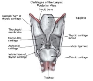
The medical information on this site is provided as an information resource only and is not to be used or relied on for any diagnostic or treatment purposes. Disarticulated cartilages left and intrinsic muscles right There are nine cartilages three unpaired and three paired 3 pairs6 that support the mammalian larynx and form its skeleton. Hindgut labeled at lower left.
The larynx also forms part of the upper respiratory tract. Larynx anterior view The larynx is a complex hollow structure located in the anterior midline region of the neckIt is anterior to the esophagus and at the level of the third to the sixth cervical vertebrae in its normal position. October 28 2021 Reading time.
Profile view of a human embryo estimated at twenty or twenty-one days old. Cartilages of the larynx Author. Arytenoid cartilage mounted on the cricoid ring with a functioning recurrent laryngeal nerve and lateral and posterior cricoarytenoid muscles form the cricoarytenoid unit a basic functional unit critical to phonatory and sphincteric functions of the larynx.
Inferiorly it continues as the cervical trachea.

1 5 Posterior View Of Larynx Showing Aryepiglottic And Oblique Download Scientific Diagram

Schematic Of The Human Larynx Framework Based On Gray 6 A Download Scientific Diagram

Cartilages Of The Larynx Posterior View Diagram Quizlet

Muscles Of Larynx Posterior View Stock Photo Alamy
Arytenoid Subluxation Practice Essentials History Of The Procedure Problem

Larynx Posterior View Diagram Quizlet

Larynx Contemporary Health Issues

Posterior View Of Larynx Human Body Vocabulary Medical Knowledge Medical Anatomy
0 comments
Post a Comment